Dual antiplatelet therapy for acute coronary syndromes: How long to continue?
www.ccjm.org
Sept. 26, 2026, 12:25 p.m.
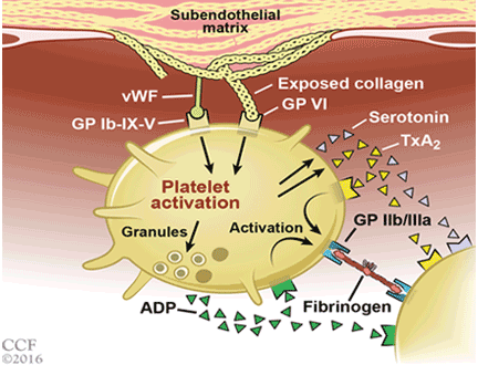
For patients with an acute coronary syndrome event, current guidelines recommend dual antiplatelet therapy for at least 12 months after drug-eluting stent placement. However, several clinical trials have assessed whether continuing dual antiplatelet therapy beyond 12 months is beneficial. We review the pros and cons of extending dual antiplatelet therapy.
Share on
Association of a platelet function testing-guided intensified
www.frontiersin.org
Sept. 26, 2026, 4:28 a.m.
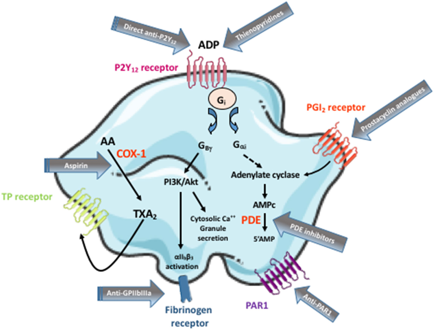
Researchers evaluated whether platelet function testing (PFT)-guided antiplatelet therapy reduces complications in patients undergoing stent-assisted coil embolization for unruptured intracranial aneurysms. The retrospective study enrolled 368 patients from two Chinese hospitals between January 2019 and September 2024, divided into a monitored group (n=219) receiving PFT-guided treatment and a non-monitored control group (n=149) receiving standard dual antiplatelet therapy (DAPT). Patients with high on-treatment platelet reactivity to clopidogrel received additional loading doses and tirofiban, while those with aspirin resistance received increased aspirin doses. The monitored strategy significantly reduced seven-day thromboembolic events (3.20% versus 8.05%, p=0.048) and thirty-day thromboembolic events (adjusted HR=0.412, p=0.028) compared to standard DAPT, with no increased bleeding complications. These findings suggest personalized, testing-guided antiplatelet intensification substantially improves safety outcomes in endovascular aneurysm treatment by identifying and managing platelet resistance, which has important clinical implications for optimizing perioperative antiplatelet management in neurovascular procedures.
Share on
Transvenous Embolization for Complex Anterior Cranial Fossa Dural Arteriovenous Fistula Following Prior Embolization: A Case Report
www.jstage.jst.go.jp
Sept. 26, 2026, 4:27 a.m.
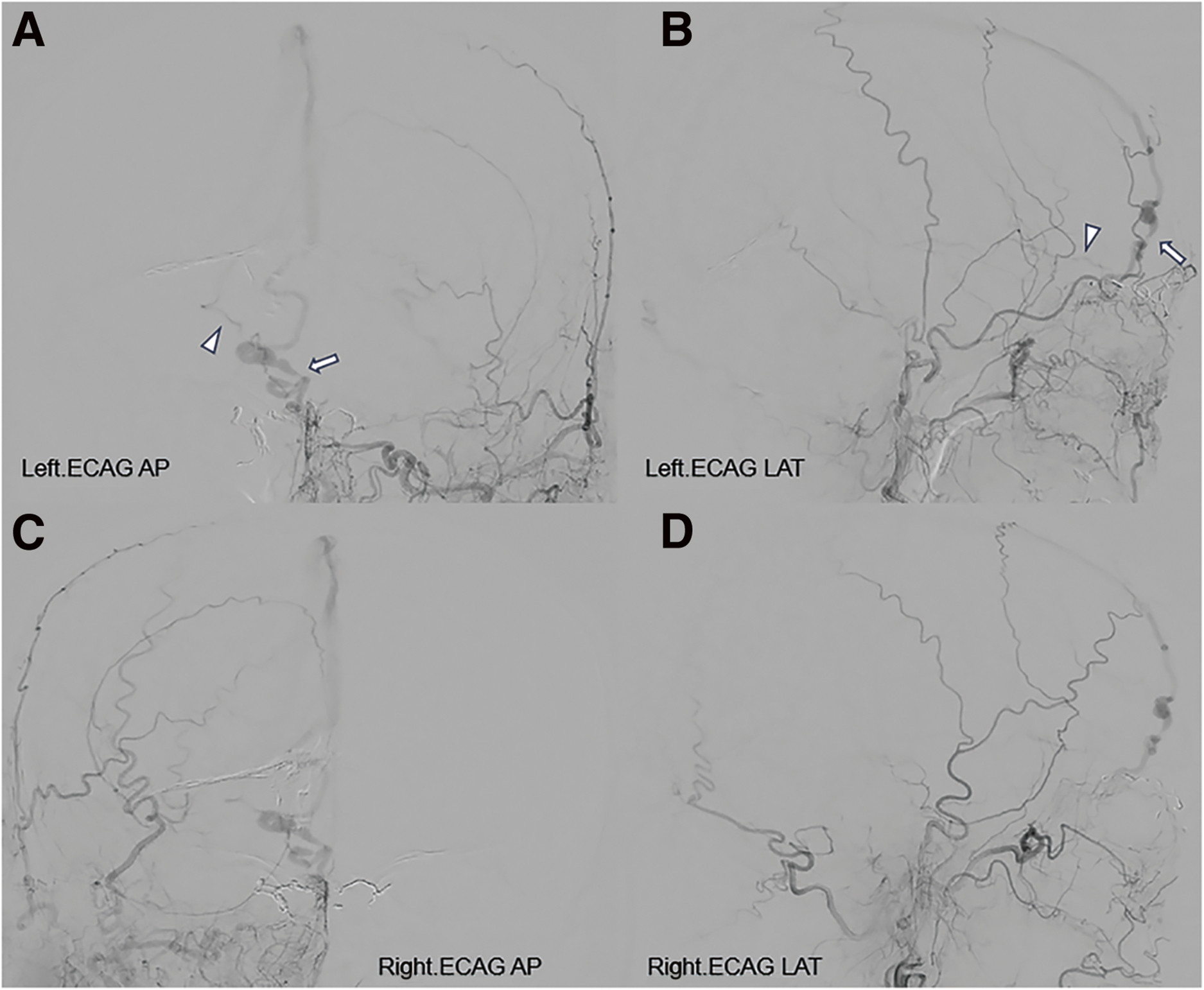
A 67-year-old patient with a history of cerebellar infarction and multiple dural arteriovenous fistulas (DAVFs) presented with recurrent headache following prior treatments. The anterior cranial fossa DAVF had persisted despite previous transarterial embolization (TAE) via bilateral ophthalmic arteries using Onyx (Medtronic). Given the lesion's location, previous arterial treatment, and the patient's strong refusal of cranial surgery, the treatment team selected transvenous embolization (TVE) as an alternative approach. This technique involved accessing the dilated draining vein directly through the venous system rather than pursuing additional arterial intervention or surgical intervention via craniotomy. The procedure was performed under general anesthesia using standard endovascular techniques, with access gained through the right internal jugular vein. TVE offered a less invasive option compared to surgical vein interruption while addressing the limitations of repeat arterial approaches. This case demonstrates how TVE can serve as a viable alternative for complex anterior cranial fossa DAVFs when conventional arterial embolization fails and patients decline surgical intervention, though the procedure carries specific risks including venous perforation, Onyx reflux, and catheter complications.
Share on
Clinical Implementation and Performance of Real-Time AI System for Device Tracking in Neurointervention: First U.S. Evaluation of Neuro-Vascular Assist - Clinical Neuroradiology
link.springer.com
Sept. 26, 2026, 4:27 a.m.
# Summary
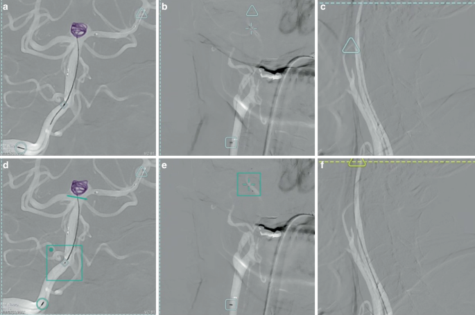
A landmark U.S. clinical evaluation assessed the real-time artificial intelligence system for device tracking during neurointerventional procedures, representing the first formal implementation study of this technology in American clinical practice. The research evaluated an AI-driven tracking system designed to monitor and guide medical devices through neuro-vascular pathways in real-time during interventional procedures. The study measured system performance across multiple clinical metrics, including tracking accuracy, procedural efficiency, and safety parameters during actual patient treatments. Key findings demonstrated the system's capability to enhance real-time visualization and device navigation while maintaining procedural safety standards. This evaluation matters significantly for the neurointerventional field because accurate device tracking can reduce procedure times, minimize radiation exposure, and potentially improve patient outcomes during complex vascular interventions. The successful implementation represents an important validation of AI-assisted technology in clinical neurovascular practice, establishing a framework for broader adoption of real-time tracking systems in interventional radiology. These results advance the integration of artificial intelligence into precision medicine within neurovascular procedures.
Share on
[PDF] Endovascular thrombectomy for mild vertebrobasilar artery occlusion
www.medrxiv.org
Sept. 23, 2026, 1:25 p.m.
# Endovascular Thrombectomy for Mild Vertebrobasilar Artery Occlusion
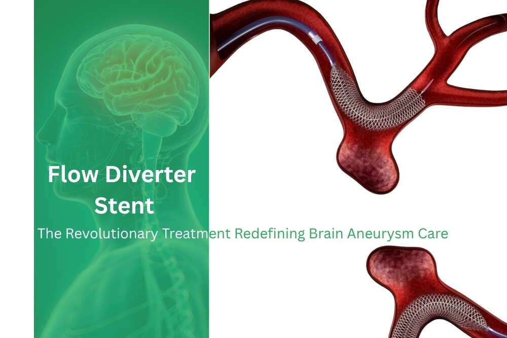
Endovascular thrombectomy, a minimally invasive interventional procedure, has emerged as a treatment approach for patients experiencing mild vertebrobasilar artery occlusion, a serious condition affecting blood flow to the brain stem and cerebellum. The vertebrobasilar system comprises the vertebral and basilar arteries, critical vessels supplying posterior circulation to the brain. While severe basilar artery occlusions typically warrant immediate mechanical thrombectomy, the optimal management strategy for mild occlusions has remained unclear due to limited clinical evidence.
This research examines the efficacy and safety of endovascular thrombectomy in mild vertebrobasilar occlusion cases, where symptoms may be less catastrophic but still significant. The procedure involves deploying specialized devices to mechanically retrieve blood clots, restoring vessel patency and cerebral perfusion. Key findings demonstrate that endovascular intervention can achieve substantial recanalization rates while maintaining acceptable safety profiles with minimal complications.
This work matters because it addresses a treatment gap for mild vertebrobasilar occlusions, potentially expanding appropriate indications for thrombectomy beyond severe cases and improving clinical outcomes for patients at risk of stroke progression or recurrent cerebrovascular events.
Share on

Antiplatelet Effects in Neurointervention: IA vs CAS
www.omnicuris.com
Sept. 23, 2026, 1:24 p.m.
Dual antiplatelet therapy with aspirin and P2Y12 inhibitors remains essential in endovascular neurointervention to prevent thromboembolic complications. However, recent comparative clinical evidence reveals that patient responses vary significantly depending on underlying vascular pathology. Intracranial aneurysms and cerebral artery stenosis represent distinct disease entities requiring differentiated treatment approaches. Aneurysms develop from hemodynamic wall shear stress and structural degradation, while cerebral artery stenosis results from atherosclerotic plaque accumulation and chronic endothelial dysfunction. These pathophysiological differences create unique microenvironments affecting platelet behavior differently. Stenotic lesions with exposed subendothelial collagen exhibit higher baseline platelet reactivity, whereas aneurysmal walls present stasis-prone outpouches where flow dynamics predominate. When neurovascular stents or flow diverters are deployed, they introduce foreign surfaces into non-atherosclerotic vessels, further distinguishing treatment responses. Although both conditions achieve similar thrombotic protection with dual antiplatelet therapy, their bleeding profiles differ substantially. Understanding these distinct safety margins enables interventional teams to refine individualized treatment regimens and optimize post-procedural recovery outcomes.
Share on
A Multicenter Prospective Study of New Flow Diverter Stent on the Treatment of Intracranial Aneurysms in China (APRESENT)
ctv.veeva.com
Sept. 23, 2026, 1:24 p.m.
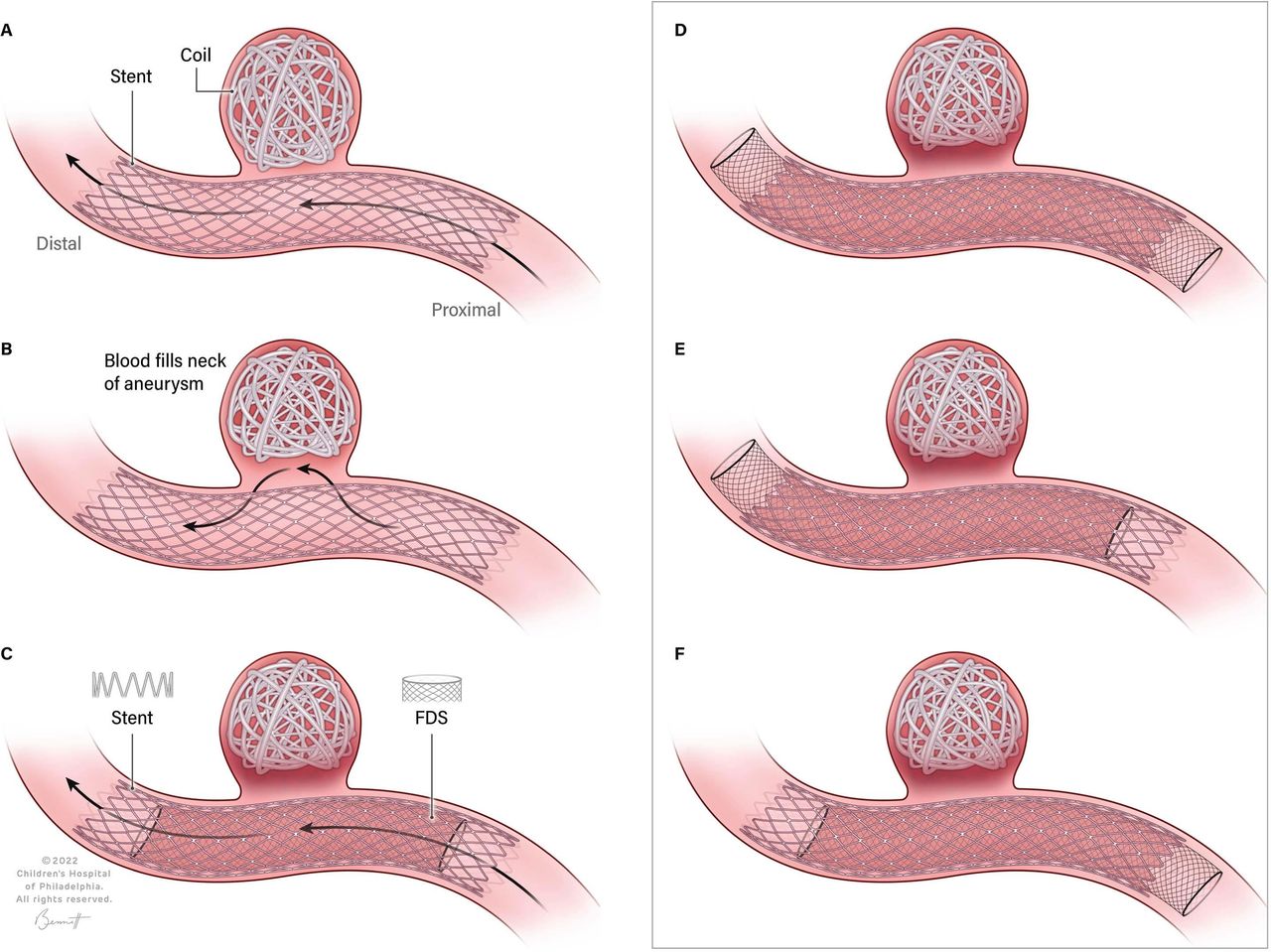
This multicenter prospective study evaluates a novel flow diverter stent for treating intracranial aneurysms, establishing rigorous inclusion and exclusion criteria to ensure patient safety and data validity. The study excludes patients with recent intracranial implants, prior endovascular interventions within 30 days, significant functional impairment (modified Rankin Scale score ≥3), or contraindications to dual antiplatelet therapy. Additional exclusion criteria encompass pre-existing stents in the parent artery, high neurological event risk, pregnancy status, and allergies to materials including platinum and cobalt-chromium alloys commonly used in flow diverter construction. By carefully controlling for these factors, the research aims to establish the safety and efficacy profile of this new stent technology. Flow diverter stents represent an important therapeutic advancement for complex aneurysm management, offering an alternative to traditional coiling and clipping approaches, making this trial's rigorous methodology essential for validating the device's clinical utility.
Share on
Disease of cervical arteries: contemporary imaging, medical management and endovascular treatment
tjcvd.org
Sept. 19, 2026, 4:25 a.m.
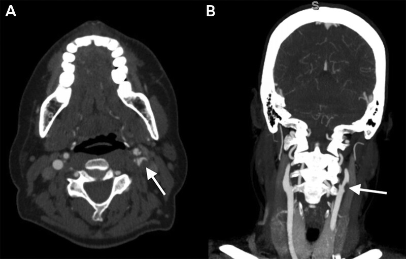
Stroke remains a leading cause of death and disability globally, with cervical artery disease accounting for a significant proportion of ischemic cerebrovascular events. Atherosclerotic narrowing of carotid and vertebral arteries causes approximately 15–20% of ischemic strokes, while non-atherosclerotic conditions such as cervical artery dissection, fibromuscular dysplasia, and large-vessel vasculitides represent important causes, particularly in younger patients. The diagnostic and therapeutic approach to cervical artery disease has transformed substantially over two decades. Historically, clinical decisions relied on luminal stenosis severity based on landmark trials like NASCET and ECST; however, emerging evidence demonstrates that stenosis alone inadequately predicts vascular lesion behavior. Contemporary assessment now emphasizes plaque vulnerability, intraplaque hemorrhage, fibrous cap integrity, inflammatory activity, and hemodynamic characteristics. Advanced imaging modalities including high-resolution vessel wall magnetic resonance imaging, multidetector computed tomography angiography, and contrast-enhanced magnetic resonance angiography now provide complementary biological and functional information. Artificial intelligence, radiomics, and quantitative imaging biomarkers have emerged as promising tools for improved plaque characterization. Therapeutically, management has evolved beyond revascularization to include intensive medical therapy, vascular risk factor modification, individualized antithrombotic strategies, and careful patient selection for carotid endarterectomy or stenting.
Share on
Perfuze Announces Final MARRS Results: Strong Reperfusion and First-Pass Performance in Pivotal Stroke Trial
finance.yahoo.com
Sept. 19, 2026, 4:25 a.m.
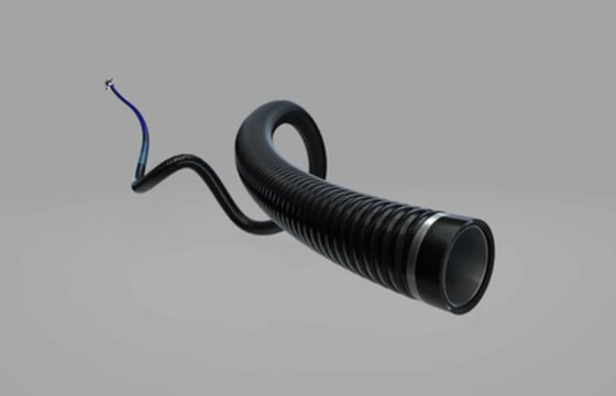
Perfuze announced final results from the MARRS pivotal study, published in the Journal of Neurointerventional Surgery, demonstrating strong efficacy for its Millipede Aspiration System in treating acute ischemic stroke. The prospective, multicenter trial enrolled 180 patients across 22 hospitals in the United States and Europe and met its primary endpoint, achieving successful reperfusion (mTICI 2b-3) in 89% of patients without rescue therapy, exceeding the prespecified performance goal. The study showed a 75% first-pass effect (mTICI 2c-3) in middle cerebral artery M1 occlusions and 96% delivery success using the Millipede88 catheter. The device's patented Corrugated™ technology enables standalone direct aspiration through a super-bore 0.088-inch catheter, eliminating the need for adjunctive devices like stent retrievers in the majority of cases. Rescue therapy was required in fewer than 8% of cases. Safety data were favorable, with a 2.3% symptomatic intracranial hemorrhage rate, no vessel perforations, and no device-related serious adverse events. These results, based on independent core laboratory adjudication, represent the highest first-pass and reperfusion rates reported in a prospective thrombectomy study and supported FDA 510(k) clearance of the Millipede88 Aspiration Catheter.
Share on
Flow diversion versus conventional endovascular treatment for ruptured basilar artery dissecting aneurysms: a single-center cohort study
www.frontiersin.org
Sept. 19, 2026, 4:24 a.m.
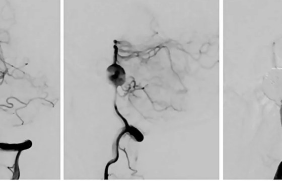
Ruptured basilar artery dissecting aneurysms (BADAs) represent rare but life-threatening conditions with elevated risks of rebleeding, brainstem ischemia, and mortality. Unlike vertebral artery dissections, the basilar trunk cannot typically be sacrificed due to its supply of critical pontine perforators, making treatment decisions particularly complex. This retrospective single-center cohort study examined 22 patients with angiographically confirmed ruptured BADA treated between February 2015 and April 2024, comparing outcomes between flow diverter (FD)-based treatment and conventional endovascular approaches. Five patients received FD-based reconstruction as primary treatment, while seventeen underwent conventional endovascular therapy. The FD group exhibited more frequent fusiform morphology (80.0% versus 23.5%) and numerically larger aneurysm diameters. At final follow-up, 80.0% of FD-treated patients achieved favorable functional outcomes (modified Rankin Scale 0–2) compared to 52.9% in the conventional group, with respective mortality rates of 20.0% and 47.1%. Although these results were not statistically significant, the findings suggest that flow diverter-based reconstruction represents a feasible option for anatomically complex or fusiform basilar dissecting aneurysms, supporting individualized treatment strategies rather than establishing clear superiority of either approach.
Share on
Signal Transduction Pathways Mediated by PECAM-1
www.ahajournals.org
Sept. 18, 2026, 10:09 a.m.
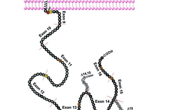
Recent studies of platelet endothelial cell adhesion molecule-1 (PECAM-1 [CD31])-deficient mice have revealed that this molecule plays an important role in controlling the activation and survival of cells on which it is expressed. In this review, we focus on the complex cytoplasmic domain of PECAM-1 and describe what is presently known about its structure, posttranslational modifications, and binding partners. In addition, we summarize findings that implicate PECAM-1 as an inhibitor of cellular activation via protein tyrosine kinase–dependent signaling pathways, an activator of integrins, and a suppressor of cell death via pathways that depend on damage to the mitochondria. The challenge of future research will be to bridge our understanding of the functional and biochemical properties of PECAM-1 by establishing mechanistic links between signals transduced by the PECAM-1 cytoplasmic domain and discrete cellular responses.
Share on
Platelet signaling: a complex interplay between inhibitory and activatory networks
www.jthjournal.org
Sept. 18, 2026, 10:01 a.m.
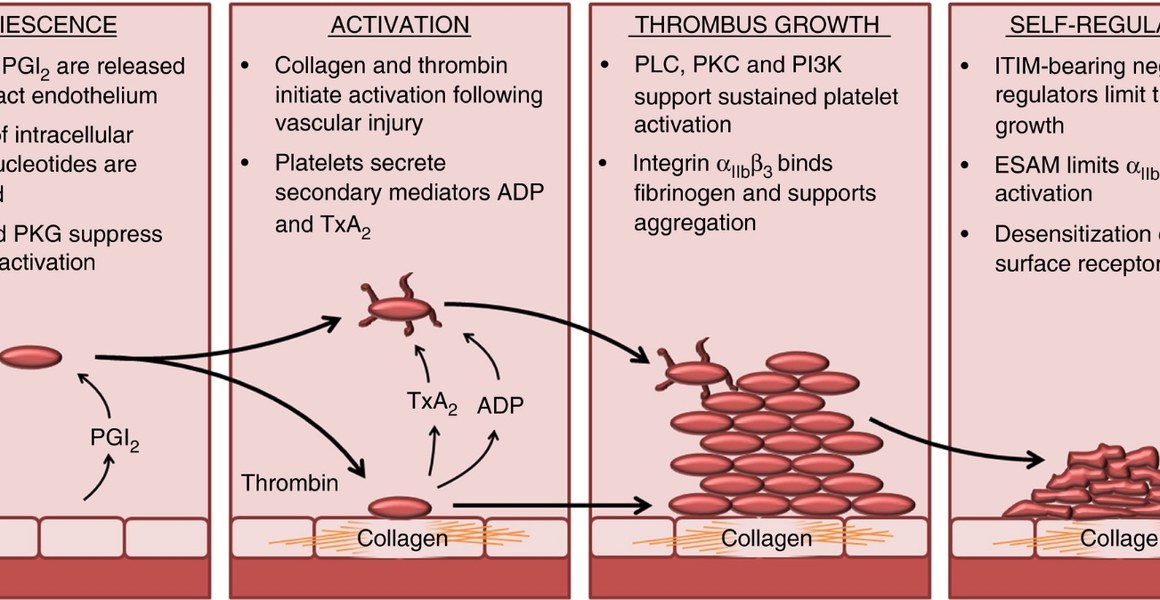
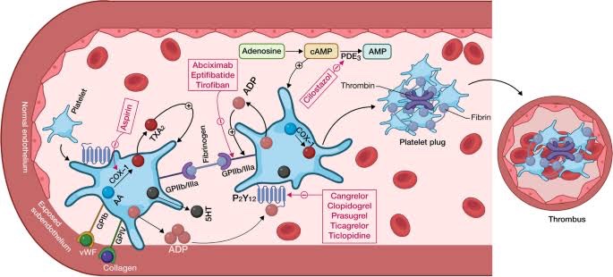
The role of platelets in hemostasis and thrombosis is dependent on a complex balance of activatory and inhibitory signaling pathways. Inhibitory signals released from the healthy vasculature suppress platelet activation in the absence of platelet receptor agonists. Activatory signals present at a site of injury initiate platelet activation and thrombus formation; subsequently, endogenous negative signaling regulators dampen activatory signals to control thrombus growth. Understanding the complex interplay between activatory and inhibitory signaling networks is an emerging challenge in the study of platelet biology, and necessitates a systematic approach to utilize experimental data effectively. In this review, we will explore the key points of platelet regulation and signaling that maintain platelets in a resting state, mediate activation to elicit thrombus formation, or provide negative feedback. Platelet signaling will be described in terms of key signaling molecules that are common to the pathways activated by platelet agonists and can be described as regulatory nodes for both positive and negative regulators.
Share on
Targeting platelet inhibition receptors for novel therapies: PECAM-1 and G6b-B
www.tandfonline.com
Sept. 18, 2026, 10 a.m.
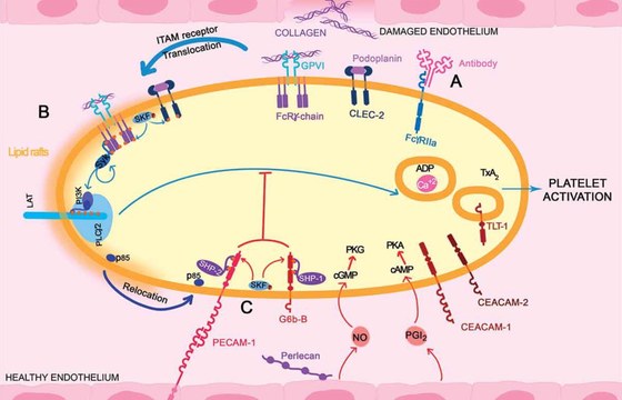
While current oral antiplatelet therapies benefit many patients, they deregulate the hemostatic balance leaving patients at risk of systemic side-effects such as hemorrhage. Dual antiplatelet treatment is the standard approach, combining aspirin with P2Y12 blockers. These therapies mainly target autocrine activation mechanisms (TxA2, ADP) and, more recently, the use of thrombin or thrombin receptor antagonists have been added to the available approaches. Recent efforts to develop new classes of anti-platelet drugs have begun to focus on primary platelet activation pathways such as through the immunoreceptor tyrosine-based activation motif (ITAM)-containing collagen receptor GPVI/FcRγ-chain complex. There are already encouraging results from targeting GPVI, with reduced aggregation and smaller arterial thrombi, without major bleeding complications, likely due to overlapping activation signaling pathways with other receptors such as the GPIb–V–IX complex. An alternative approach to reduce platelet activation could be to inhibit this signaling pathway by targeting the inhibitory pathways intrinsic to platelets. Stimulation of endogenous negative modulators could provide more specific inhibition of platelet function, but is this feasible? In this review, we explore the potential of the two major platelet immunoreceptor tyrosine-based inhibitory motif (ITIM)-containing inhibitory receptors, G6b-B and PECAM-1, as antithrombotic targets.
Share on
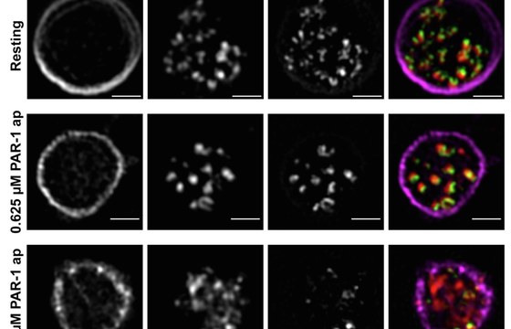
Quantitative super-resolution imaging of platelet degranulation reveals differential release of von Willebrand factor and von Willebrand factor propeptide from alpha-granules
www.jthjournal.org
Sept. 16, 2026, 2:37 p.m.
Von Willebrand factor (VWF) and VWF propeptide (VWFpp) are stored in eccentric nanodomains within platelet alpha-granules. VWF and VWFpp can undergo differential secretion following Weibel-Palade body exocytosis in endothelial cells; however, it is unclear if the same process occurs during platelet alpha-granule exocytosis. Using a high-throughput 3-dimensional super-resolution imaging workflow for quantification of individual platelet alpha-granule cargo, we studied alpha-granule cargo release in response to different physiological stimuli.
Share on

Brain Aneurysm Surgery: Clipping vs Coiling Explained
acibademinternational.com
Sept. 16, 2026, 1:32 p.m.
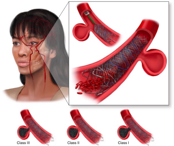
Brain aneurysms are weak, bulging spots in brain artery walls that risk rupture and life-threatening subarachnoid hemorrhage. Two primary treatment approaches exist: surgical clipping, where neurosurgeons place a small metal clip across the aneurysm through a skull opening, and endovascular coiling, which fills the aneurysm with soft coils delivered via catheter through groin or wrist arteries. Both methods are employed for ruptured aneurysms as emergency treatment and for selected unruptured aneurysms with significant bleeding risk. Newer techniques include flow diverters—stent-like mesh tubes redirecting blood flow—and stent-assisted coiling for specific aneurysm configurations. Treatment decisions depend on aneurysm characteristics including size, location, shape, and neck width, alongside patient age and health status. Not all aneurysms require intervention; many small incidental findings never cause symptoms. Multidisciplinary teams comprising neurosurgeons, neuroradiologists, and neurologists determine appropriate treatment by weighing aneurysm rupture risk against procedural risks. Detailed imaging including CT scans and angiograms guides treatment planning and selection.
Share on
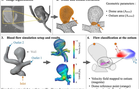
Clinical outcome prediction of Contour Neurovascular System treatment based on pre-treatment geometric and hemodynamic characteristics
link.springer.com
Sept. 16, 2026, 1:31 p.m.
Researchers have evaluated the Contour Neurovascular System, a medical device used in neurovascular interventions, by analyzing how pre-treatment geometric and hemodynamic characteristics can predict clinical outcomes. The study examined patient data to identify which structural and blood flow parameters measured before treatment were most predictive of post-intervention success. By applying computational analysis to anatomical and hemodynamic features—such as vessel dimensions, flow patterns, and lesion geometry—the investigators developed predictive models to forecast treatment efficacy. These findings matter significantly for clinical practice, as they enable physicians to better identify which patients will likely benefit most from Contour Neurovascular System treatment before proceeding with intervention. This approach supports personalized medicine by allowing clinicians to optimize patient selection and potentially improve overall outcomes while reducing unnecessary procedures for unsuitable candidates. The research contributes valuable insights to interventional neurology and vascular surgery planning.
Share on
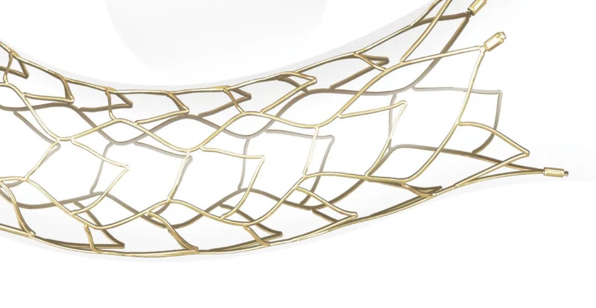
Small Coating, Big Ambition: NeuroVita's SECURA™ Self-Expandable Intracranial Stent Launches With AlchiMedics’ eG™ NTMA Coating Technology
mp.weixin.qq.com
Sept. 16, 2026, 11:28 a.m.
NeuroVita has announced the Chinese regulatory approval and commercial launch of SECURA™, a coated intracranial stent system designed to assist in treating wide-neck intracranial aneurysms.
SECURA combines low metal coverage with eG™ NTMA, a coating technology invented and developed by AlchiMedics, intended to reduce platelet adhesion and thrombosis risk. Its optimised “dragon-scale” mesh promotes vessel-wall apposition and stable support in complex anatomy. Compatibility with 0.017-inch and 0.021-inch microcatheters, together with tipped and tipless configurations, provides flexibility across different access routes and procedural needs.
The device complements NeuroVita’s AUCURA™ flow-diverting stent, expanding the company’s portfolio for intracranial aneurysm treatment.
Share on
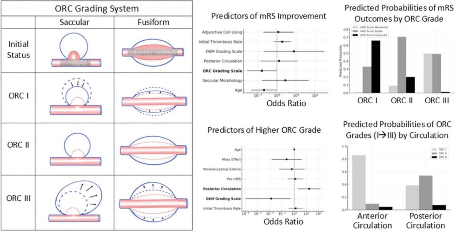
Follow-up Results of Partially Thrombosed Intracranial Aneurysms with Cross-sectional Imaging: The Case for Introducing a New Grading System
www.ajnr.org
Sept. 12, 2026, 11 a.m.
The CSI-based 3-group categorization provides a more clinically meaningful assessment of PTIAs treated with FDs than conventional angiographic classifications, because it uniquely captures size changes of the thrombosed mass and correlates with functional outcomes.
Share on
Antiplatelets and antithrombotics in neurointerventional procedures: Guideline update
jnis.bmj.com
Sept. 12, 2026, 10:59 a.m.
Although the quality of evidence is lower than for coronary interventions due to a lower number of patients and procedures, neurointerventional antiplatelet and antithrombotic management shares several themes. Prospective and randomized studies are needed to strengthen the data supporting these recommendations.
Share on
Flow diverter stents for endovascular treatment of aneurysms: a comparative study of efficacy and safety between FREDX and FRED
jnis.bmj.com
Sept. 12, 2026, 10:57 a.m.
The FREDX device shows improved complete occlusion rates at 1 year compared with the FRED device while maintaining a favourable safety profile, indicating its potential advantage in the treatment of cerebral aneurysms.
Share on